
ARTICULAR THE FIBROUS CONNECTIVE TISSUE CARTILAGE DEFECTS
A 20 years' girl studying in engineering, arrived to us for her acutely locked & unpredictable knee. In a recent randomized controlled trial, comparing ACI versus microfracture Saris et al. 56 reported better results in chondral defects treated with characterized autologous chondrocytes than microfracture group, revealed simply arthryl a alkohol by better structural repair examined by histomorphometry and general histological evaluation. Though the clinical scores were comparable in both groups, cells regeneration was superior in ACI group. Long-term follow-up is again required to ascertain the survival of the ACI graft.
Although the fibrous connective tissue cartilage appears relatively uniform on gross inspection, the positioning of collagen and the focus of water, chondrocytes, and proteoglycans varies across the density of the tissue 13, 15 The presence of layers in cartilage viewed on MR images implies a correlation with histologic zonal organization. Rubenstein ainsi que al. 16 established a link between tissue composition and the MR graphic by demonstrating T2 anisotropy within cartilage. In their very own study, the pattern of layering seen on MR images varied as the fibrous connective tissue cartilage orientation relative to B0 changed, proving that the influence of structural components within cartilage was responsible for the presence of layering.A great articular cartilage injury, or perhaps chondral injury, may happen as a result of a pivot or angle on a bent knee, a direct blow to the knee, or damage because a patient gets older. Sometimes, chondral injuries may accompany an injury to a ligament including the anterior cruciate ligament (ACL). Tiny 4 flex silver skład pieces of the anudar cartilage can break away and float around in the knee as loose bodies, causing locking, capturing and/or swelling. For just about all patients, there is zero clear history reported of a single injury. This type of injury may result from a series of minor injuries that have occurred over time.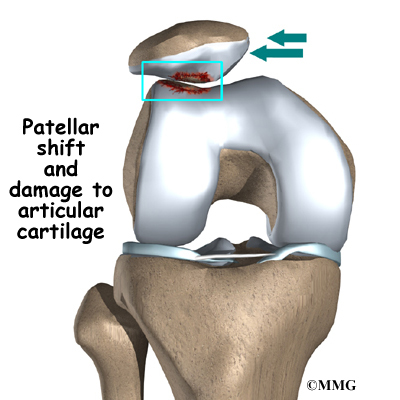 Figure 2. The catabolic physiology top rated to articular cartilage breakdown. Interleukin-1 is among the principle cytokines that initiates a chute that leads to chondrocyte cell death and extracellular matrix breakdown. NSAIDs inhibit collaflex 60 prostaglandins, such as PGE2, from stimulating chondrocyte GENETICS matrix synthesis thereby adding to articular cartilage degeneration.Three-dimensional bSSFP imaging depicts fluid with increased signal strength while preserving the sign intensity of cartilage, offering excellent synovial fluid-cartilage contrast. With recent advances on gradient coils, shorter TEs have become possible, helping remove off-resonance artifacts due to field inhomogeneity, such while banding artifacts. Yet , these previously common artifacts happen to be still a problem with the use of great field strength (3 T) or a long TR. Just because a short TR does not allow sufficient image resolution for cartilage imaging, multiple acquisitions can be needed to achieve higher resolution.